
• Illustrations
  • animations
  • 3d models
Pricing
  • Illustrations
  • Specialized Illustrations
  • Disease and Pathology Illustrations
accepted payments

Contact

support@sciepro.comhttps://sciepro.com
SciePro Distribution GmbH
Zum Exerzierhaus 15
14469 Potsdam
Germany

Legal

Legal noticeTerms and conditionsPrivacy policyPricing

Company

About usBlogContactFAQ

© Copyright 2026 | SciePro Distribution GmbH

Disease and Pathology Illustrations

Medically reviewed illustrations and 3D animations of diseases and pathological changes.

Recent search :

Hero banner
Established Malignant Growth (Descending Colon)

Established Malignant Growth (Descending Colon)

Rectal Cancer, a Type of Colorectal Cancer

Rectal Cancer, a Type of Colorectal Cancer

Malignant Rectal Neoplasm, Gross Anatomy

Malignant Rectal Neoplasm, Gross Anatomy

Colorectal Cancer Positioned in the Sigmoid Colon

Colorectal Cancer Positioned in the Sigmoid Colon

Cancerous Growth Located (Sigmoid Colon)

Cancerous Growth Located (Sigmoid Colon)

Colorectal Cancer (Transverse Colon)

Colorectal Cancer (Transverse Colon)

Identification of a Malignant Tumor in the Transverse Colon

Identification of a Malignant Tumor in the Transverse Colon

Solid Fibroadenoma Tumor Located (Breast)

Solid Fibroadenoma Tumor Located (Breast)

Fibrotic Malignant Tumor in the Breast

Fibrotic Malignant Tumor in the Breast

Acute Hematoma Found (Breast)

Acute Hematoma Found (Breast)

Painful Abscess (Breast)

Painful Abscess (Breast)

Malignant Tumor Affecting a Breast Lymph Node

Malignant Tumor Affecting a Breast Lymph Node

Stage 0 Non-invasive Breast Neoplasm

Stage 0 Non-invasive Breast Neoplasm

Stage 1 Malignant Breast Tumor

Stage 1 Malignant Breast Tumor

Characteristics of a Stage 2 Breast Cancer

Characteristics of a Stage 2 Breast Cancer

Stage 3 Advanced Malignant Breast Neoplasm

Stage 3 Advanced Malignant Breast Neoplasm

Stage 4 Metastatic Breast Cancer

Stage 4 Metastatic Breast Cancer

Multiple Benign Cysts Located (Breast)

Multiple Benign Cysts Located (Breast)

Breast Exhibiting Symptoms of Mastitis

Breast Exhibiting Symptoms of Mastitis

Area of Tissue Necrosis (Breast)

Area of Tissue Necrosis (Breast)

Benign Tumor Known as a Breast Fibroadenoma

Benign Tumor Known as a Breast Fibroadenoma

Dense, Malignant Fibrotic Cancer

Dense, Malignant Fibrotic Cancer

Localized Malignant Tumor of the Breast

Localized Malignant Tumor of the Breast

Earliest Stage of Breast Cancer Identified as Stage 0

Earliest Stage of Breast Cancer Identified as Stage 0

Malignant Tumor Classified as Breast Cancer Stage 1

Malignant Tumor Classified as Breast Cancer Stage 1

Development of Breast Cancer Categorized as Stage 2

Development of Breast Cancer Categorized as Stage 2

Advanced Breast Cancer Diagnosis at Stage 3

Advanced Breast Cancer Diagnosis at Stage 3

Metastatic Breast Cancer Classified as Stage 4

Metastatic Breast Cancer Classified as Stage 4

Appearance of Multiple Fluid-filled Breast Cysts

Appearance of Multiple Fluid-filled Breast Cysts

Mammary Glands Affected by Fibrocystic Changes

Mammary Glands Affected by Fibrocystic Changes

  • Previous
  • 1
  • 2
  • 3
  • ...
  • 19
  • ...
  • 33
  • 34
  • 35
  • Next

Disease and Pathology Illustrations and Animations

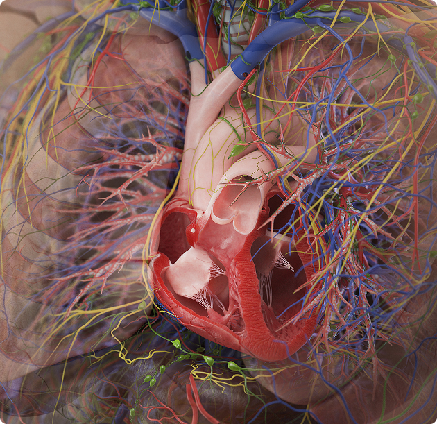
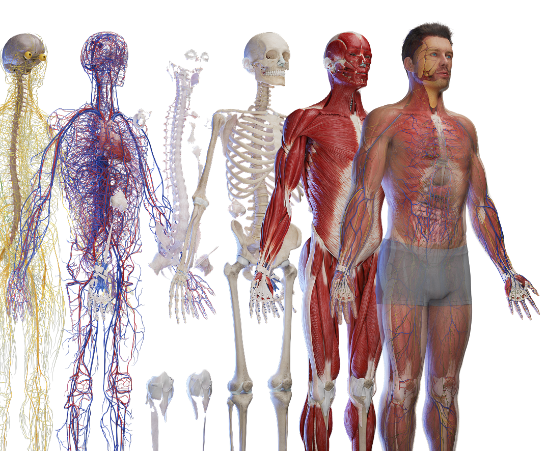
The disease-specific category covers pathology across the organ systems. Assets depict the affected structures and the changes that define each condition, including cardiovascular, respiratory, digestive, neurological, musculoskeletal, and other system pathologies, shown against normal anatomy for comparison where useful. Because the affected structures are rendered from the same labeled 3D model used for healthy anatomy across the library, pathology stays consistent with normal anatomy and disease progression can be shown as a sequence. Every asset is reviewed by our Medical Advisory Board. The collection supports clinical and patient education, pharmaceutical content, continuing medical education, and editorial use. Assets are available as high-resolution illustrations and 3D animations, licensed under Educational, Standard, or Extended terms through a subscription with credits or as individual purchases.
About category image

Why Choose SciePro Illustrations

Professional-grade medical visuals trusted by leading institutions

icon

Verified by Medical Experts

Every illustration is reviewed by our Medical Advisory Board and aligned with the Terminologia Anatomica, the international standard for anatomical nomenclature. Accuracy is the baseline for every asset in the library.

icon

Exclusive to SciePro

Every asset in our library is produced in-house and licensed exclusively through this platform. You will not find our illustrations on Shutterstock, Getty Images, or any other agency.

icon

Rooted in 20 Years of Practice

Since 2005, our work has been licensed more than 5 million times and used by institutions including the Mayo Clinic, Merck, and Arthrex. The library reflects two decades of production experience in medical visualization.

Show all illustrations
Human anatomy models

Frequently Asked Questions

Pathology across organ systems, depicting the affected structures and the changes that define each condition, often shown against normal anatomy.

Yes. Because pathology is built on the same anatomy model, progression can be shown as a sequence of stages.

Yes. Every asset is reviewed by our Medical Advisory Board.

High-resolution JPG and PNG, with layered PSD on selected assets, and MP4 for animations.

Yes. Educational, Standard, and Extended licenses are available. See https://library.sciepro.com for details.

Need Help Choosing?

Our team can help you find the right plan and license for your specific use case.

Human anatomy models

Testimonials

Jeremy London

Jeremy London

MD, Board Certified Cardiovascular Surgeon

starstarstarstarstar

“It was a pleasure and honor to work with SciePro. They make the best anatomical animations I have seen on the internet. Very easy to work with and hope to work with them again in the future. I would highly recommend SciePro to anyone!”

Linda Peterson

Linda Peterson

MD, Professor of Medicine at Washington University School of Medicine in cardiology

starstarstarstarstar

“I have been teaching school children of all ages about heart health and disease in February, heart month, since 2016, and I am so excited to use the exceptional images from Sciepro.com. The images are stunningly realistic and accurate and really help me to teach about heart health. They also show how the heart relates to its neighboring organs, and there are images that depict the initiation and progression of atherosclerosis.”

Dr. Nathan Berner

Dr. Nathan Berner

Board Certified Orthospinology Practitioner

starstarstarstarstar

“The animations are the best out there. I use models for education related to our business and these are top notch!”

Dr. Christian Rivera

Dr. Christian Rivera

Plastic and Reconstructive Surgeon

starstarstarstarstar

“As a healthcare professional, I fully trust SciePro GmbH for accurate and high-quality medical illustrations. Their ability to transform complex medical concepts into clear and detailed images is unmatched, making communication with both colleagues and patients easier. These illustrations not only enhance understanding but are also essential tools in medical education and treatment planning. SciePro GmbH demonstrates a deep understanding of the medical field and a commitment to excellence.”

Trusted by leading institutions worldwide

University of Calgary
Stratasys
DocCheck
Med Graz
Vantari VR
Augmed
Hite Vision
IMRA
AnthroTek
Allrise
Johnson and Johnson
Novartis
P&G
Pfizer
Publicis
St. Jude Medical
Stanford University