• Illustrations
  • animations
  • 3d models
Pricing
  • Illustrations
  • Musculoskeletal System
  • Muscle Anatomy Illustrations
accepted payments

Contact

support@sciepro.comhttps://sciepro.com
SciePro Distribution GmbH
Zum Exerzierhaus 15
14469 Potsdam
Germany

Legal

Legal noticeTerms and conditionsPrivacy policyPricing

Company

About usBlogContactFAQ

© Copyright 2026 | SciePro Distribution GmbH

Muscle Anatomy Illustrations

Medically reviewed illustrations and 3D animations of the skeletal muscles by region.

Recent search :

Hero banner
Posterior Perspective of the Trapezius Muscle

Posterior Perspective of the Trapezius Muscle

Triceps Under the Skin, Posterior View

Triceps Under the Skin, Posterior View

Triceps, Posterior View

Triceps, Posterior View

Latissimus Dorsi Under the Skin, Posterior View

Latissimus Dorsi Under the Skin, Posterior View

Latissimus Dorsi, Posterior View

Latissimus Dorsi, Posterior View

Anterior Perspective of the Muscles of the Lower Arm Under the Skin

Anterior Perspective of the Muscles of the Lower Arm Under the Skin

Brachioradialis Under the Skin, Anterior View

Brachioradialis Under the Skin, Anterior View

Brachioradialis, Anterior View

Brachioradialis, Anterior View

Flexor Carpi Radialis Under the Skin, Anterior View

Flexor Carpi Radialis Under the Skin, Anterior View

Flexor Carpi Radialis, Anterior View

Flexor Carpi Radialis, Anterior View

Flexor Digitorum Profundus Under the Skin, Anterior View

Flexor Digitorum Profundus Under the Skin, Anterior View

Flexor Digitorum Profundus, Anterior View

Flexor Digitorum Profundus, Anterior View

Flexor Digitorum Superficialis Under the Skin, Anterior View

Flexor Digitorum Superficialis Under the Skin, Anterior View

Flexor Digitorum Superficialis, Anterior View

Flexor Digitorum Superficialis, Anterior View

Flexor Pollicis Longus Under the Skin, Anterior View

Flexor Pollicis Longus Under the Skin, Anterior View

Flexor Pollicis Longus, Anterior View

Flexor Pollicis Longus, Anterior View

Oblique Cord Under the Skin, Anterior View

Oblique Cord Under the Skin, Anterior View

Oblique Cord, Anterior View

Oblique Cord, Anterior View

Anterior Perspective of the Palmaris Longus Under the Skin

Anterior Perspective of the Palmaris Longus Under the Skin

Palmaris Longus, Anterior View

Palmaris Longus, Anterior View

Anterior Perspective of the Pronator Teres Under the Skin

Anterior Perspective of the Pronator Teres Under the Skin

Pronator Teres, Anterior View

Pronator Teres, Anterior View

Lateral Perspective of the Muscles of the Lower Arm Under the Skin

Lateral Perspective of the Muscles of the Lower Arm Under the Skin

Abductor Pollicis Longus Under the Skin, Lateral View

Abductor Pollicis Longus Under the Skin, Lateral View

Abductor Pollicis Longus, Lateral View

Abductor Pollicis Longus, Lateral View

Lateral Perspective of the Anconeus Under the Skin

Lateral Perspective of the Anconeus Under the Skin

Anconeus, Lateral View

Anconeus, Lateral View

Brachioradialis Under the Skin, Lateral View

Brachioradialis Under the Skin, Lateral View

Brachioradialis, Lateral View

Brachioradialis, Lateral View

Extensor Carpi Radialis Brevis Under the Skin, Lateral View

Extensor Carpi Radialis Brevis Under the Skin, Lateral View

  • Previous
  • 1
  • 2
  • 3
  • ...
  • 40
  • ...
  • 127
  • 128
  • 129
  • Next

Muscular System and Muscle Anatomy Illustrations and Animations

The muscular system category covers the skeletal muscles of the body. Assets include the muscles of the head and neck, trunk, and upper and lower limbs, organized by region and muscle group, with their origins, insertions, and associated tendons. Superficial and deep muscle layers are shown separately so individual muscles can be identified in context. Structures are available in anterior, posterior, lateral, and medial views, as isolated muscles, regional muscle groups, and the full muscle figure, with bone references where helpful for origins and insertions. Every asset is rendered from one labeled 3D model built to Terminologia Anatomica and reviewed by our Medical Advisory Board, so muscles stay registered with the underlying skeleton. The collection supports anatomy, sports medicine, and physiotherapy education, clinical communication, medical device content, and editorial use. Assets are available as high-resolution illustrations and 3D animations, licensed under Educational, Standard, or Extended terms through a subscription with credits or as individual purchases.
About category image

Why Choose SciePro Illustrations

Professional-grade medical visuals trusted by leading institutions

icon

Verified by Medical Experts

Every illustration is reviewed by our Medical Advisory Board and aligned with the Terminologia Anatomica, the international standard for anatomical nomenclature. Accuracy is the baseline for every asset in the library.

icon

Exclusive to SciePro

Every asset in our library is produced in-house and licensed exclusively through this platform. You will not find our illustrations on Shutterstock, Getty Images, or any other agency.

icon

Rooted in 20 Years of Practice

Since 2005, our work has been licensed more than 5 million times and used by institutions including the Mayo Clinic, Merck, and Arthrex. The library reflects two decades of production experience in medical visualization.

Show all illustrations
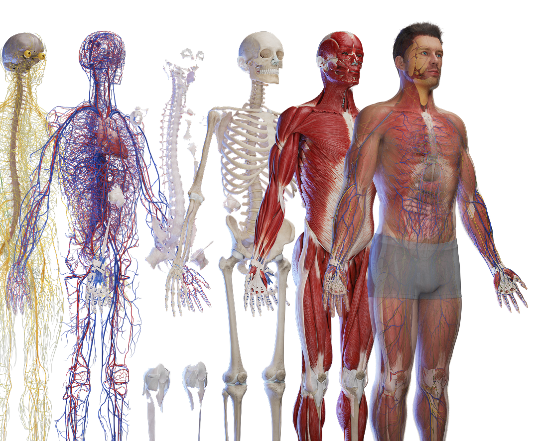
Human anatomy models

Frequently Asked Questions

The skeletal muscles by body region and group, with origins, insertions, tendons, and separate superficial and deep layers.

Yes. Layered assets let you isolate superficial or deep muscles from the same registered model.

Yes. Every asset follows Terminologia Anatomica nomenclature and is reviewed by our Medical Advisory Board.

High-resolution JPG and PNG, with layered PSD on selected assets, and MP4 for animations.

Yes. Educational, Standard, and Extended licenses are available. See https://library.sciepro.com for details.

Need Help Choosing?

Our team can help you find the right plan and license for your specific use case.

Human anatomy models

Testimonials

Jeremy London

Jeremy London

MD, Board Certified Cardiovascular Surgeon

starstarstarstarstar

“It was a pleasure and honor to work with SciePro. They make the best anatomical animations I have seen on the internet. Very easy to work with and hope to work with them again in the future. I would highly recommend SciePro to anyone!”

Linda Peterson

Linda Peterson

MD, Professor of Medicine at Washington University School of Medicine in cardiology

starstarstarstarstar

“I have been teaching school children of all ages about heart health and disease in February, heart month, since 2016, and I am so excited to use the exceptional images from Sciepro.com. The images are stunningly realistic and accurate and really help me to teach about heart health. They also show how the heart relates to its neighboring organs, and there are images that depict the initiation and progression of atherosclerosis.”

Dr. Nathan Berner

Dr. Nathan Berner

Board Certified Orthospinology Practitioner

starstarstarstarstar

“The animations are the best out there. I use models for education related to our business and these are top notch!”

Dr. Christian Rivera

Dr. Christian Rivera

Plastic and Reconstructive Surgeon

starstarstarstarstar

“As a healthcare professional, I fully trust SciePro GmbH for accurate and high-quality medical illustrations. Their ability to transform complex medical concepts into clear and detailed images is unmatched, making communication with both colleagues and patients easier. These illustrations not only enhance understanding but are also essential tools in medical education and treatment planning. SciePro GmbH demonstrates a deep understanding of the medical field and a commitment to excellence.”

Trusted by leading institutions worldwide

University of Calgary
Stratasys
DocCheck
Med Graz
Vantari VR
Augmed
Hite Vision
IMRA
AnthroTek
Allrise
Johnson and Johnson
Novartis
P&G
Pfizer
Publicis
St. Jude Medical
Stanford University